3D virtual reconstruction of the Kebara 2 Neandertal thorax
Asier Gomez-Olivencia, Alon Barash, Daniel Garcia-Martinez, Mikel Arlegi, Patricia Kramer, Markus Bastir, Ella Been - Dept. Estratigrafia y Paleontologia, Facultad de Ciencia y Tecnologia, Universidad del Pais Vasco/Euskal Herriko Unibertsitatea (UPV/EHU), Bilbao, Spain; IKERBASQUE. Basque Foundation for Science, Spain; Equipe de Paleontologie Humaine, CNRS, Departement de Prehistoire, Museum National d’Histoire naturelle, Musee de l’Homme, Paris, France; Centro Mixto UCMISCIII de Evolucion y Comportamiento Humanos, Madrid, Spain; Faculty of Medicine in the Galilee, Bar-Ilan University, Zefat, Israel; Paleoanthropology Group, Museo Nacional de Ciencias Naturales (CSIC), Madrid, Spain; Universite de Bordeaux, Pessac, France; Departments of Anthropology and Orthopaedics and Sports Medicine, University of Washington, Seattle, USA; Department of Sports Therapy, Faculty of Health Professions, Ono Academic College, Kiryat Ono, Israel; Department of Anatomy and Anthropology, Sackler Faculty of Medicine, Tel Aviv University, Israel.
![3D virtual reconstruction of the Kebara 2 Neandertal thorax]()
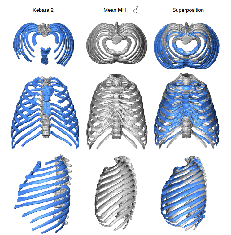
The size and shape of the Neandertal thorax has been debated since the first discovery of Neandertal ribs more than 150 years ago, with workers proposing different interpretations ranging from a Neandertal thoracic morphology that is indistinguishable from modern humans, to one that was significantly different from them. Here, we provide a virtual 3D reconstruction of the thorax of the adult male Kebara 2 Neandertal. Our analyses reveal that the Kebara 2 thorax is significantly different but not larger from that of modern humans, wider in its lower segment, which parallels his wide bi-iliac breadth, and with a more invaginated vertebral column. Kinematic analyses show that rib cages that are wider in their lower segment produce greater overall size increments (respiratory capacity) during inspiration. We hypothesize that Neandertals may have had a subtle, but somewhat different breathing mechanism compared to modern humans.