In situ observation of mechanical damage within a SiC-SiC ceramic matrix composite
L. Saucedo-Mora, T. Lowe, S. Zhao, P.D. Lee, P.M. Mummery, T.J. Marrow - Institute Eduardo Torroja for Construction Sciences-CSIC, Madrid, Spain ; Department of Materials, University of Oxford, UK ; Manchester X-ray Imaging Facility, The University of Manchester, UK ; Research Complex at Harwell, Rutherford Appleton Laboratory, UK ; School of Mechanical, Aerospace and Civil Engineering, The University of Manchester, UK
SiC-SiC ceramic matrix composites are candidate materials for fuel cladding in Generation IV nuclear fission reactors and as accident tolerant fuel clad in current generation plant.
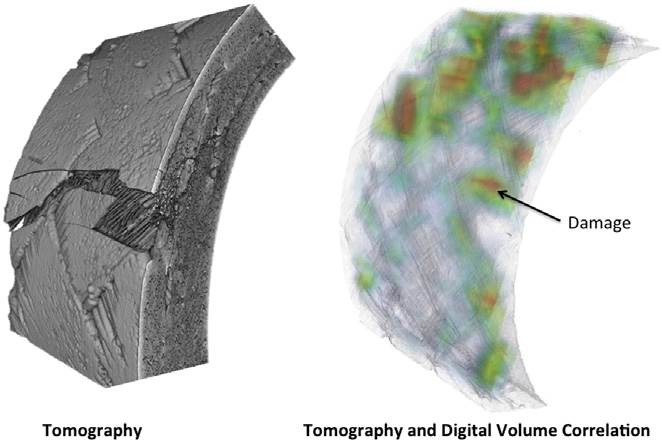
Experimental methods are needed that can detect and quantify the development of mechanical damage, to support modelling and qualification tests for these critical components. In situ observations of damage development have been obtained of tensile and C-ring mechanical test specimens of a braided nuclear grade SiC-SiC ceramic composite tube, using a combination of ex situ and in situ computed X-ray tomography observation and digital volume correlation analysis. The gradual development of damage by matrix cracking and also the influence of non-uniform loading are examined.
How Amira-Avizo Software is used
Segmentation of the porosity via image intensity thresholding was done using the Avizo (Fig. 2e). The contrast between pores and solid material is sufficiently high that the quantitative analysis is insensitive to the chosen segmentation thresholds. The analysis of the segmented pores shows that tomographs at different resolutions captured an equivalent total porosity (Fig. 3a). The average porosity content, measured within segments of 0.015 mm length, varies along the tube section and similar data for the average porosity (~5e7%) are obtained in medium resolution (17 mm voxel) and high resolution (3.25 mm voxel) tomography scans that studied different tubes. The smallest pores, which make up a small fraction of the total porosity, are best characterised at higher resolution (Fig. 3b); the lowest resolution tomographs examined a sufficiently large volume to observe the largest pores in the population, which are up to 1mmin size when measured in the tangential direction of each layer of the tube.