Microstructural analysis of TRISO particles using multi-scale X-ray computed tomography
TRISO particles, a composite nuclear fuel built up by ceramic and graphitic layers, have outstanding high temperature resistance. TRISO fuel is the key technology for High Temperature Reactors (HTRs) and the Generation IV Very High Temperature Reactor (VHTR) variant.
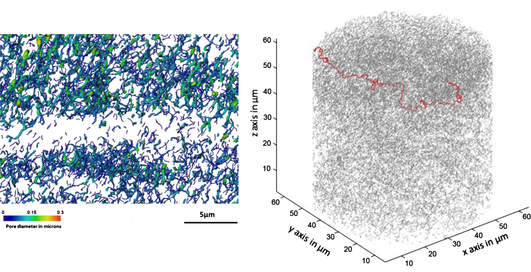
TRISO offers unparalleled containment of fission products and is extremely robust during accident conditions. An understanding of the thermal performance and mechanical properties of TRISO fuel requires a detailed knowledge of pore sizes, their
distribution and interconnectivity. Here 50 nm, nano-, and 1 lm resolution, micro-computed tomography (CT), have been used to quantify non-destructively porosity of a surrogate TRISO particle
at the 0.3–10 lm and 3–100 lm scales respectively. This indicates that pore distributions can reliably be measured down to a size approximately 3 times the pixel size which is consistent with the segmentation process. Direct comparison with Scanning Electron Microscopy (SEM) sections indicates that
destructive sectioning can introduce significant levels of coarse damage, especially in the pyrolytic carbon layers. Further comparative work is required to identify means of minimizing such damage for SEM studies. Finally since it is non-destructive, multi-scale time-lapse X-ray CT opens the possibility
of intermittently tracking the degradation of TRISO structure under thermal cycles or radiation conditions in order to validate models of degradation such as kernel movement. X-ray CT in-situ experimentation of TRISO particles under load and temperature could also be used to understand the internal changes that occur in the particles under accident conditions.