Operative Anatomy of the Human Skull: A Virtual Reality Expedition
Benjamin K Hendricks, MD Akash J Patel, MD Jerome Hartman Mark F Seifert, PhD Aaron Cohen-Gadol, MD, MSc, MBA - Goodman Campbell Brain and Spine and Indiana University School of Medicine, Indianapolis, Indiana
![Operative Anatomy of the Human Skull: A Virtual Reality Expedition]()
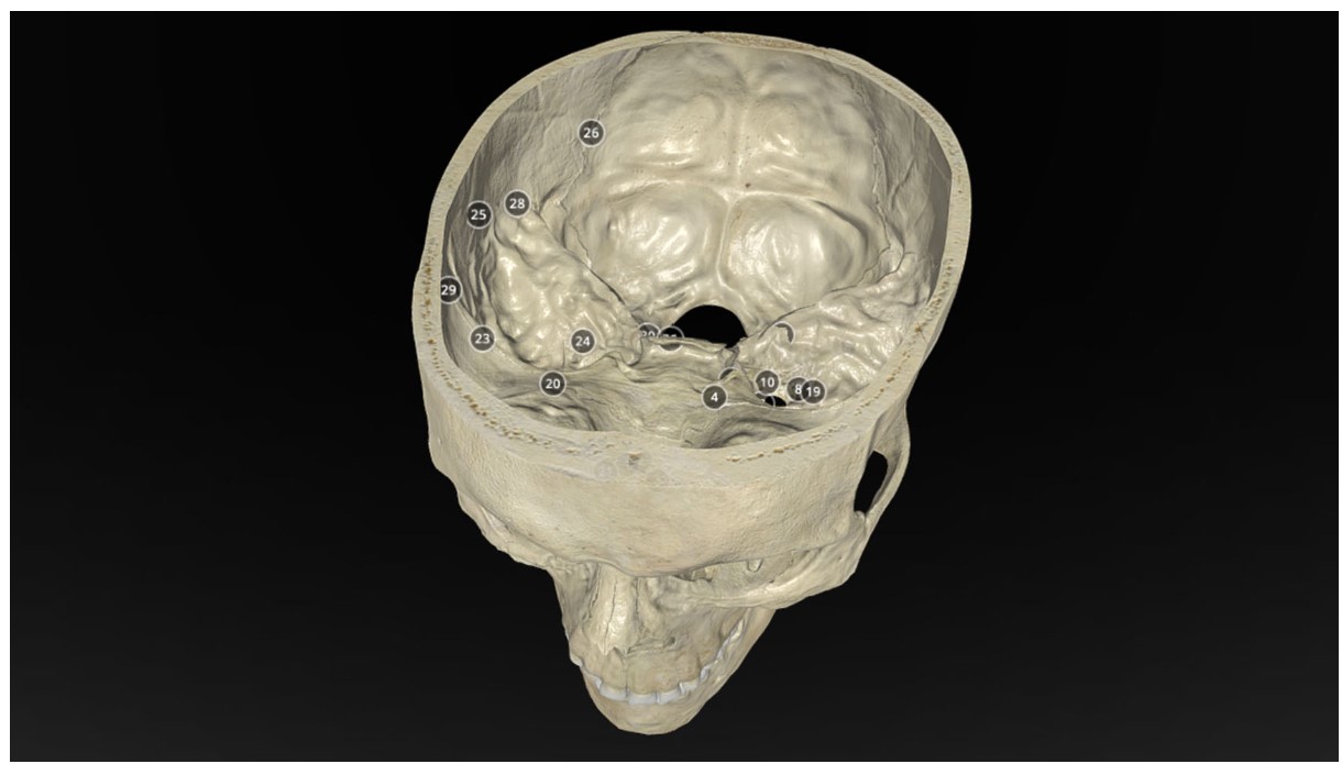
The human cranial vault possesses an incredible, complex anatomical intricacy. Bridging the divide between 2-dimensional (2D) learning resources and the 3-dimensional (3D) world in which the anatomy becomes clinically relevant poses an intellectual challenge. Advances in computer graphics and modelling technologies have allowed increasingly accurate and representative resources to supplement cadaveric dissection specimens. To create accurate virtual models of all cranial bones to augment education, research, and clinical endeavours. Through a careful analysis of osteological specimens and high-resolution radiographic studies, a highly accurate virtual model of the human skull was created and annotated with relevant anatomical landmarks. The skull was divided into 6 major segments including frontal, ethmoid, sphenoid, temporal, parietal, and occipital bones. These bones were thoroughly annotated to demonstrate the intricate anatomical features.