Ptychographic X-ray CT characterization of the osteocyte lacuno-canalicular network in a male rat's glucocorticoid induced osteoporosis model
Antonia Ciani, Hechmi Toumi, Stéphane Pallu, Esther H.R.Tsai, Ana Diaz, Manuel Guizar-Sicairos, Mirko Holler, Eric Lespessailles, Cameron M.Kewish | Synchrotron Soleil, France
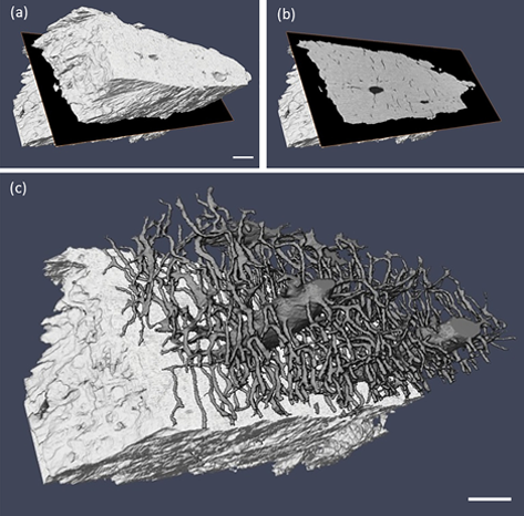
Ptychographic X-ray computed tomography (PXCT) is a quantitative imaging modality that non-destructively maps the 3D electron density inside an object with tens of nanometers spatial resolution. This method provides unique access to the morphology and structure of the osteocyte lacuno-canalicular network (LCN) and nanoscale density of the tissue in the vicinity of an osteocyte lacuna. Our findings indicate that PXCT can non-destructively provide detailed, nanoscale information on the 3D organization of the LCN in correlative studies of pathologies, such as osteoporosis, leading to improved diagnosis and therapy.