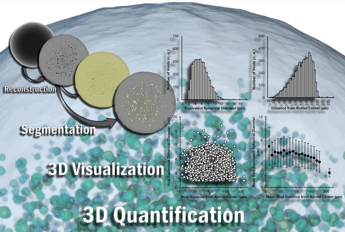
Seeing through nuclear fuel: Three-dimensional, nondestructive X-ray microscopy and volumetric analyses of neutron-irradiated TRISO-coated fuel kernels
The three-dimensional (3D) characterization of nuclear fuel with X-ray microscopy has historically proven difficult, due to uranium’s high attenuation of easily accessible X-rays, both in a laboratory setting and at a synchrotron user facility. However, this imaging modality provides nondestructive information that can be used to investigate morphological changes arising from external stimuli (e.g., neutron irradiation, high-temperature testing).

Using an appropriate X-ray energy spectrum and an adequate X-ray filter, suitable transmissions through properly sized nuclear fuel specimens can be achieved. Here, we present the methods and results of using a commercially available, laboratory-based X-ray microscope (XRM) to examine the extent of 3D morphological changes of tristructural isotropic (TRISO)-coated fuel particles, specifically uranium oxide/uranium carbide fuel kernels, after high-temperature neutron irradiation.