The visible skeleton 2.0: phenotyping of cartilage and bone in fixed vertebrate embryos and foetuses based on X-ray microCT
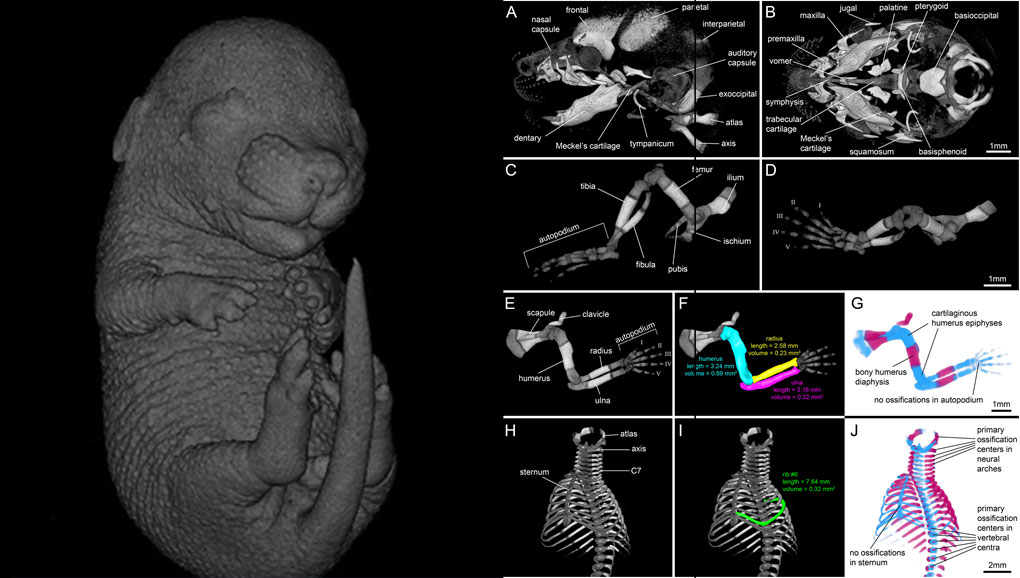
For decades, clearing and staining with Alcian Blue and Alizarin Red has been the gold standard to image vertebrate skeletal development. Here, we present an alternate approach to visualise bone and cartilage based on X-ray microCT imaging, which allows the collection of genuine 3D data of the entire developing skeleton at micron resolution.

Our novel protocol is based on ethanol fixation and staining with Ruthenium Red, and efficiently contrasts cartilage matrix, as demonstrated in whole E16.5 mouse foetuses and limbs of E14 chicken embryos. Bone mineral is well preserved during staining, thus the entire embryonic skeleton can be imaged at high contrast. Differences in X-ray attenuation of ruthenium and calcium enable the spectral separation of cartilage matrix and bone by dual energy microCT (microDECT). Clearing of specimens is not required. The protocol is simple and reproducible. We demonstrate that cartilage contrast in E16.5 mouse foetuses is adequate for fast visual phenotyping. Morphometric skeletal parameters are easily extracted. We consider the presented workflow to be a powerful and versatile extension to the toolkit currently available for qualitative and quantitative phenotyping of vertebrate skeletal development.