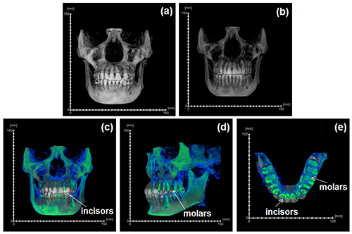
UCSF School of Dentistry uses Avizo software to extract changes in periodontal ligament space and tooth movement due to orthodontic therapy
During the acts of biting and chewing, the muscles of the jaw (consisting of the masseter, temporalis, and medial pterygoid in the elevator group, and lateral pterygoid as the main depressor) generate forces that dictate jaw kinematics . The movement of jaws hinges about the temporomandibular joint and are brought together by the muscles attached to respective bones through bone-tendon interfaces known as entheses . Thus, chewing forces affect aspects of craniofacial structure as well as bone formation and resorption.
The insertion of orthodontic braces changes the forces acting at the cementum-ligament and bone-ligament entheses, and induces orthodontic tooth movement. In this article, we analyze physical movement of teeth due to orthodontic braces commonly placed in humans to facilitate favorable occlusion.
Articulation of mandibular and maxillary jaws and interdigitation of respective teeth of a teenage patient before and after orthodontic intervention for a year was visualized using cone beam computed tomography (CBCT) at the UCSF School of Dentistry. Prior to analysis of the CBCTs, voxel size was calibrated (0.25 μm 3 per voxel). Centers of rotation of CBCT data sets from before and after intervention were registered using the Register Image function in Avizo software to identify gross tooth movement in the mandibular and maxillary jaws of the patient.