University of Washington researchers use Amira software towards aiding the pathology of pancreatic cancer
For nearly 100 years, pathology for cancer diagnosis has involved a standard, but complex series of steps to process tissue biopsies procured from a patient in the clinic. Many procedures are a direct result of the fact that observation and evaluation of specimens by pathologists occur using a standard microscope (in 2D).
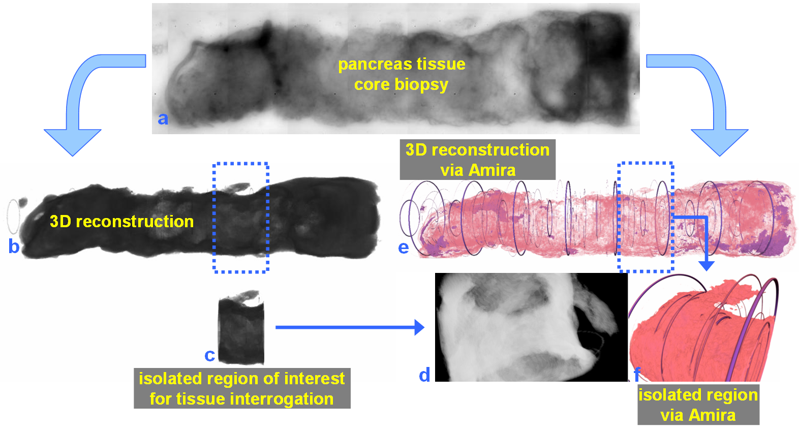
In 2014, the Human Photonics Laboratory at the University of Washington demonstrated that the rudimentary operations of a pathology laboratory may be replicated on whole, unsectioned tissue biopsies using microfluidics within a credit card-sized device . The device may potentially reduce the entire pathology laboratory’s infrastructure and physical footprint for 3D optical imaging of tissue biopsies . Imaging tissue biopsies (in 3D) makes utility of the entire patient specimen prior to sectioning, provides a fundamental gain in optical (diagnostic) data, and may permit initial biopsy triage and evaluation within the workflow of traditional pathology.