Welcome to the Amira-Avizo Software Use Case Gallery
Below you will find a collection of use cases of our 3D data visualization and analysis software. These use cases include scientific publications, articles, papers, posters, presentations or even videos that show how Amira-Avizo Software is used to address various scientific and industrial research topics.
Use the Domain selector to filter by main application area, and use the Search box to enter keywords related to specific topics you are interested in.

Regional Aneurysm Wall Enhancement is Affected by Local Hemodynamics: A 7T MRI Study
Aneurysm wall enhancement has been proposed as a biomarker for inflammation and instability. However, the mechanisms of aneurysm wall enhancement remain unclear. We used 7T MR imaging to determine the effect of flow in different regions of the wall.
Twenty-three intracranial aneurysms imaged with 7T MR imaging and 3D angiography were studied with computational fluid dynamics. Local flow conditions were compared between aneurysm wall enhancement and nonenhanced regions. Aneurysm wall en... Read more
S. Hadad, F. Mut, B.J. Chung, J.A. Roa, A.M. Robertson, D.M. Hasan, E.A. Samaniego and J.R. Cebral

Serial block-face electron microscopy (SBEM) provides nanoscale 3D ultrastructure of embedded and stained cells and tissues in volumes of up to 107 µm3. In SBEM, electrons with 1–3 keV energies are incident on a specimen block, from which backscattered electron (BSE) images are collected with x, y resolution of 5–10 nm in the block-face plane, and successive layers are removed by an in situ ultramicrotome. Sp... Read more
Q. He, M. Hsueh, G. Zhang, D. C. Joy & R. D. Leapman

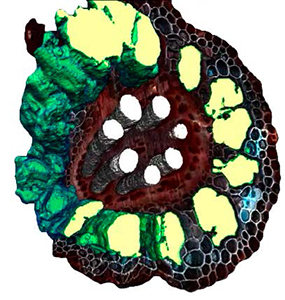
L4iS uses Avizo software to analyze 3D images from laser ablation tomography
Lasers for Innovative Solutions, LLC (L4iS) is developing a new class of tomography technology with the aim of allowing material characterization in three dimensions with sub-micron resolution. The method uses a nanosecond, Q-switched, pulsed ultraviolet laser coupled with high-resolution imaging to generate highly detailed specimen models. Using this system, sequential images similar to light-sheet fluorescence microscopy are used to digitally reconstruct the specimen.
Read more
Brian Reinhardt and Benjamin Hall, L4iS (USA)