4D imaging of polymer electrolyte membrane fuel cell catalyst layers by soft X-ray spectro-tomography

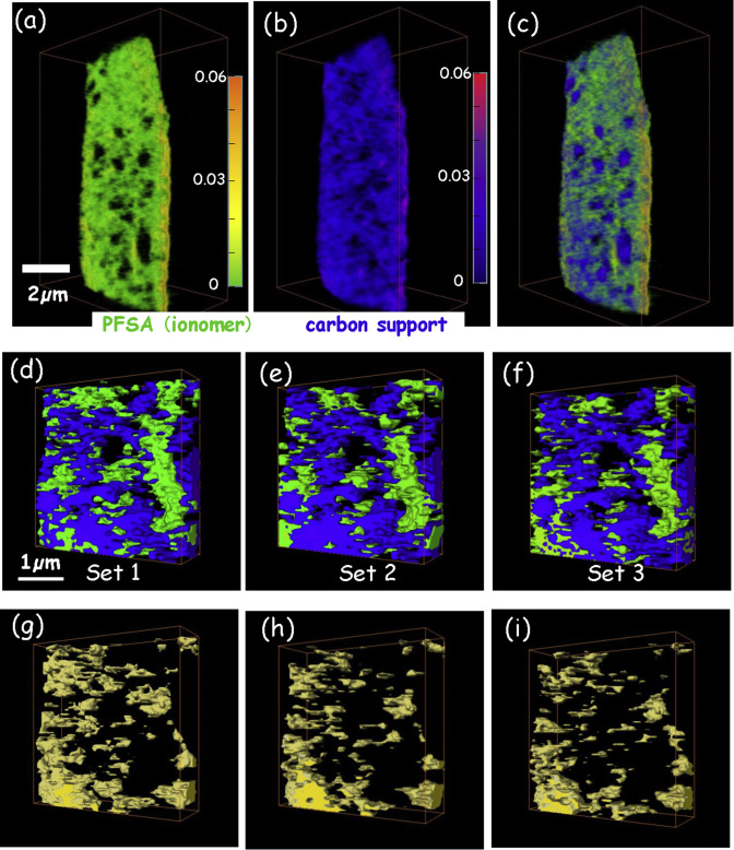
4D imaging – the three-dimensional distributions of chemical species determined using multi-energy X-ray tomography – of cathode catalyst layers of polymer electrolyte membrane fuel cells (PEM-FC) has been measured by scanning transmission x-ray microscopy (STXM) spectro-tomography at the C 1s and F 1s edges. In order to monitor the effects of radiation damage on the composition and 3D structure of the perfluorosulfonic acid (PFSA) ionomer, the same volume was measured 3 times sequentially, with spectral characterization of that same volume at several time points during the measurements. The changes in the average F 1s spectrum of the ionomer in the cathode as the measurements progressed gave insights into the degree of chemical modification, fluorine mass loss, and changes in the 3D distributions of ionomer that accompanied the spectro-tomographic measurement. The PFSA ionomer-in-cathode is modified both chemically and physically by radiation damage. The 3D volume decreases anisotropically. By reducing the incident flux, partial defocusing (50 nm spot size), limiting the number of tilt angles to 14, and using compressed sensing reconstruction, we show it is possible to reproducibly measure the 3D structure of ionomer in PEM-FC cathodes at ambient temperature while causing minimal radiation damage.