A detailed canine brain label map for neuroimaging analysis
Dogs have recently become an important model species for comparative social and cognitive neuroscience. Brain template-related label maps are essential for functional magnetic resonance imaging (fMRI) data analysis, to localize neural responses. In this study, we present a detailed, individual-based, T1-weighted MRI-based brain label map used in dog neuroimaging analysis. Methods: A typical, medium-headed dog (a 7.5-year-old
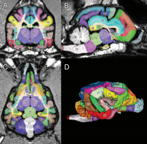
male Golden Retriever) was selected from a cohort of 22 dogs, based on brain morphology (shape, size, and gyral pattern), to serve as the template for a label map. Results: Eighty-six 3-dimensional labels were created to highlight the main cortical (cerebral gyri on the lateral and medial side) and subcortical (thalamus, caudate nucleus, amygdala, and hippocampus) structures of the prosencephalon and diencephalon, and further main parts of brainstem
(mesencephalon and rhombencephalon). Discussion: Importantly, this label map is (a) considerably more detailed than any available dog brain template; (b) it is easy to use with freeware and commercial neuroimaging software for MRI and fMRI analysis; and (c) it can be registered to other existing templates, including a recent average-based dog brain template. Using the coordinate system and label map proposed here can enhance precision and standard
localization during future canine neuroimaging studies.