Automatic method for estimation of in situ effective contact angle from X-ray micro tomography images of two-phase flow in porous media
Multiphase flow in porous media is strongly influenced by the wettability of the system, which affects the arrangement of the interfaces of different phases residing in the pores.
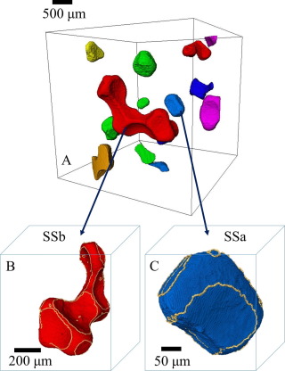
We present a method for estimating the effective contact angle, which quantifies the wettability and controls the local capillary pressure within the complex pore space of natural rock samples, based on the physical constraint of constant curvature of the interface between two fluids. This algorithm is able to extract a large number of measurements from a single rock core, resulting in a characteristic distribution of effective in situ contact angle for the system, that is modelled as a truncated Gaussian probability density distribution. The method is first validated on synthetic images, where the exact angle is known analytically; then the results obtained from measurements within the pore space of rock samples imaged at a resolution of a few microns are compared to direct manual assessment. Finally the method is applied to X-ray micro computed tomography (micro-CT) scans of two Ketton cores after waterflooding, that display water-wet and mixed-wet behaviour. The resulting distribution of in situ contact angles is characterized in terms of a mixture of truncated Gaussian densities.