Label-free 3D-CLEM using endogenous tissue landmarks
We demonstrate feasibility of the workflow by combining in vivo 2-photon microscopy and focused ion beam scanning electron microscopy (FIB/SEM) to dissect the role of astrocytic coverage in the persistence of dendritic spines.
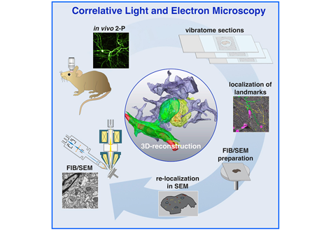
Emerging 3D correlative light and electron microscopy (CLEM) approaches enable studying neuronal structure-function relations at unprecedented depth and precision. However, established protocols for the correlation of light and electron micrographs rely on the introduction of artificial fiducial markers, such as polymer beads or near-infrared brandings, which might obscure or even damage the structure under investigation. Here, we report a general applicable ”flat embedding” preparation, enabling high-precision overlay of light and scanning electron micrographs, using exclusively endogenous landmarks in the brain: blood vessels, nuclei and myelinated axons. Furthermore, we demonstrate feasibility of the workflow by combining in vivo 2-photon microscopy and focused ion beam scanning electron microscopy (FIB/SEM) to dissect the role of astrocytic coverage in the persistence of dendritic spines.