The use of contrast enhancement techniques in X-ray imaging of lithium–ion battery electrodes
Oluwadamilola O. Taiwo , Donal P. Finegan , Jeff Gelb , Christian Holzner , Daniel J.L. Brett , Paul R. Shearing - The Electrochemical Innovation Lab, Department of Chemical Engineering, University College London, WC1E 7JE, UK | Carl Zeiss X-ray Microscopy Inc., Pleasanton, CA 94588, USA
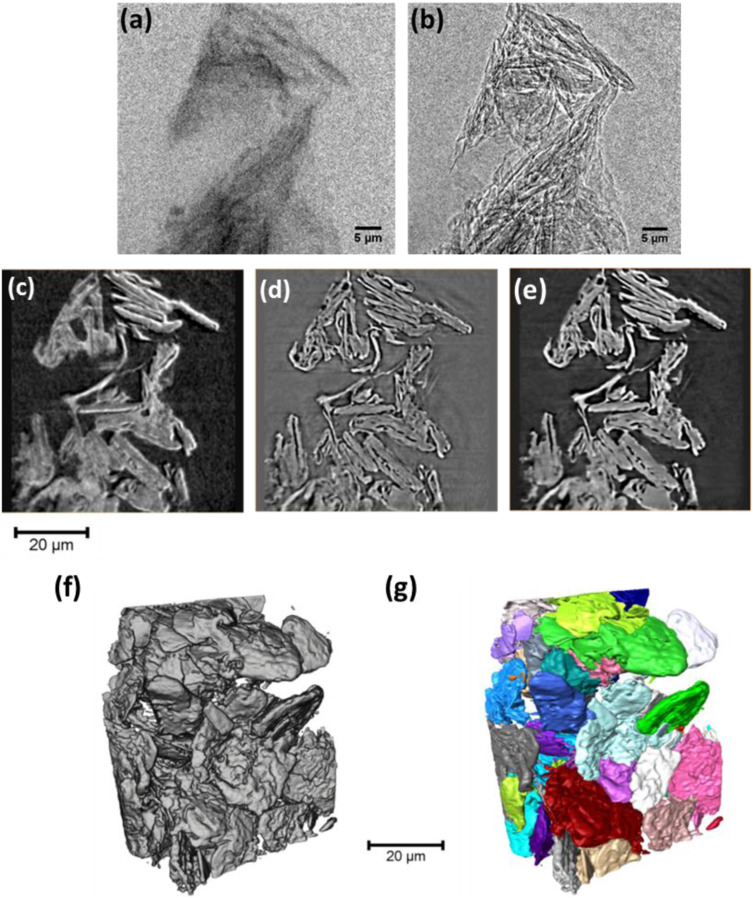
Understanding the microstructural morphology of Li–ion battery electrodes is crucial to improving the electrochemical performance of current Li–ion battery systems and in developing next-generation power systems. The use of 3D X-ray imaging techniques, which are continuously evolving, provides a noninvasive platform to study the relationship between electrode microstructure and performance at various time and length scales. In addition to characterizing a weakly (X-ray) absorbing graphite electrode at multiple length scales, we implement an approach for obtaining improved nano-scale image contrast on a laboratory X-ray microscope by combining information obtained from both absorption–contrast and Zernike phase-contrast X-ray images.