Micron-scale crack propagation in laser-irradiated enamel and dentine studied with nano-CT
Abtesam Aljdaimi, Hugh Devlin, Mark Dickinson, Timothy Burnett, Thomas J. A. Slater - Division of Dentistry, Faculty of Biology, Medicine and Health, University of Manchester, UK; College of Dentistry, Asmarya University, Zliten, Libya; Photon Science Institute, School of Physics and Astronomy, University of Manchester, UK; Henry Moseley X-ray Imaging Facility, School of Materials, University of Manchester, UK
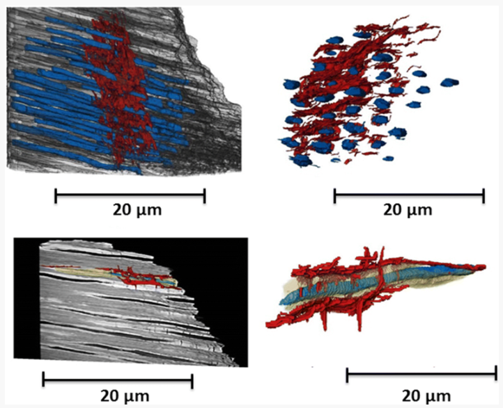
The aim of this study was to see the effect of Er:YAG laser irradiation in dentine and compare this with its effect in enamel. The mechanism of crack propagation in dentine was emphasised and its clinical implications were discussed. A possible mechanism is that laser radiation is transmitted down the dentinal tubules causing micro-cracks to form in the dentinal tubule walls that tend to be limited to this region. Crack might be a source of fracture as it represents a weak point and subsequently might lead to a failure in restorative dentistry.