Welcome to the Amira-Avizo Software Use Case Gallery
Below you will find a collection of use cases of our 3D data visualization and analysis software. These use cases include scientific publications, articles, papers, posters, presentations or even videos that show how Amira-Avizo Software is used to address various scientific and industrial research topics.
Use the Domain selector to filter by main application area, and use the Search box to enter keywords related to specific topics you are interested in.

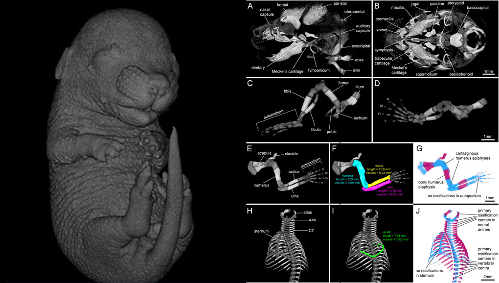
For decades, clearing and staining with Alcian Blue and Alizarin Red has been the gold standard to image vertebrate skeletal development. Here, we present an alternate approach to visualise bone and cartilage based on X-ray microCT imaging, which allows the collection of genuine 3D data of the entire developing skeleton at micron resolution.
Our novel protocol is based on ethanol fixation and staining with Ruthenium Red, and efficiently contrasts cartilage matrix, as demonstrated in wh... Read more
Simone Gabner, Peter Böck, Dieter Fink, Martin Glösmann, Stephan Handschuh

GEVES uses Avizo software to study and control plant seeds
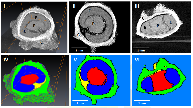
At GEVES, Microtomography (micro-CT), which is a technique using X-rays to investigate internal anatomy and morphology of organisms without destruction, is applied to plant seed quality control and phenotyping. Several applications have been developed using micro-CT coupled with Avizo software development, such as for example in the Measurements of internal and external sugar beet seed structures for seed phenotyping
Read more
Ghassen Trigui, Laurence Le Corre, Dominique Honoré and Karima Boudehri-Giresse - 2D/3D Imaging - R&D team, GEVES

L4iS uses Avizo software to analyze 3D images from laser ablation tomography
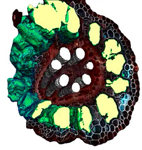
Lasers for Innovative Solutions, LLC (L4iS) is developing a new class of tomography technology with the aim of allowing material characterization in three dimensions with sub-micron resolution. The method uses a nanosecond, Q-switched, pulsed ultraviolet laser coupled with high-resolution imaging to generate highly detailed specimen models. Using this system, sequential images similar to light-sheet fluorescence microscopy are used to digitally reconstruct the specimen.
Read more
Brian Reinhardt and Benjamin Hall, L4iS (USA)