3D Failure Analysis of Pure Mechanical and Pure Chemical Degradation in Fuel Cell Membranes
Lifetime-limiting failure of fuel cell membranes is generally attributed to their chemical and/or mechanical degradation. Although both of these degradation modes occur concurrently during operational duty cycles, their uncoupled investigations can provide useful insights into their individual characteristics and consequential impacts on the overall membrane failure.
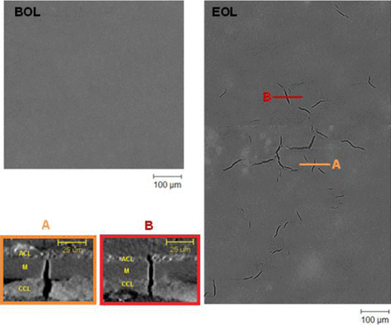
X-ray computed tomography is emerging as an advantageous tool for fuel cell failure analysis due to its non-destructive and non-invasive 3D imaging capabilities at ambient conditions. In the present work, post-mortem failure analysis of pure mechanical and pure chemical membrane degradation modes is performed three-dimensionally using this technique. A uniquely comprehensive analysis afforded by this technique reveals that membrane failure is almost exclusively characterized by crack formations during mechanical degradation and by severe thinning accompanied by electrode shorting and pinhole formation during chemical degradation, respectively. Catalyst layer cracks, particularly on the cathode side, are found to interact strongly with mechanically induced membrane cracks. The conjoint effect of chemical and mechanical stressors is established as a necessary requirement for: (i) exclusive membrane crack development independent of catalyst layer cracks; and (ii) crack branching during membrane crack propagation. Overall, the membrane failure analysis improves its reliability and quantitative character with the adoption of 3D imaging.