Juvenile Ovine Ex Vivo Larynges: Phonatory, Histologic, and Micro CT Based Anatomic Analyses
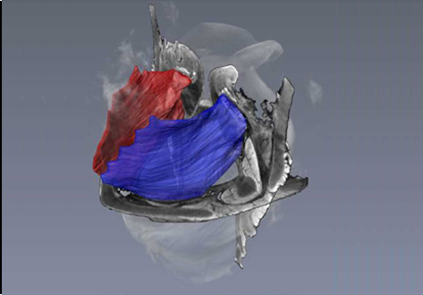
It is well known that the phonatory process changes during the life span. However, detailed investigations on potential factors concerned are rare. To deal with this issue, we performed extended biomechanical, macro anatomical, and histological analyses of the contributing laryngeal structures in ex vivo juvenile sheep models. Altogether twelve juvenile sheep larynges were analyzed within the phonatory experiments. Three different elongation levels and 16 different flow levels were applied to achieve a large variety of phonatory conditions. Vocal fold dynamics and acoustical and subglottal signals could be analyzed for 431 experimental runs. Subsequently, for six juvenile larynges microcomputed tomography following virtual 3D reconstruction was performed. The remaining six juvenile larynges as well as six ex vivo larynges from old sheep were histologically and immunohistologically analyzed. Results for juveniles showed more consistent dynamical behavior compared to old sheep larynges due to vocal fold tissue alterations during the life span. The phonatory process in juvenile sheep seems to be more effective going along with a greater dynamic range. These findings are supported by the histologically detected higher amounts of elastin and hyaluronic acid in the lamina propria of the juvenile sheep. The 3D reconstructions of the thyro-arytenoid muscles (TAM) showed a symmetrical shape. Intraindividual volume and surface differences of the TAM were small and comparable to those of aged sheep. However, TAM dimensions were statistically significant smaller for juvenile larynges. Finally, topographical landmarks were introduced for later comparison with other individuals and species. This work resulted in detailed functional, immunohistological, and anatomical information that was not yet reported. This data will also provide reference information for therapeutic strategies regarding aging effects, e.g. laryngeal muscle treatment by functional electrical stimulation.