Welcome to the Amira-Avizo Software Use Case Gallery
Below you will find a collection of use cases of our 3D data visualization and analysis software. These use cases include scientific publications, articles, papers, posters, presentations or even videos that show how Amira-Avizo Software is used to address various scientific and industrial research topics.
Use the Domain selector to filter by main application area, and use the Search box to enter keywords related to specific topics you are interested in.
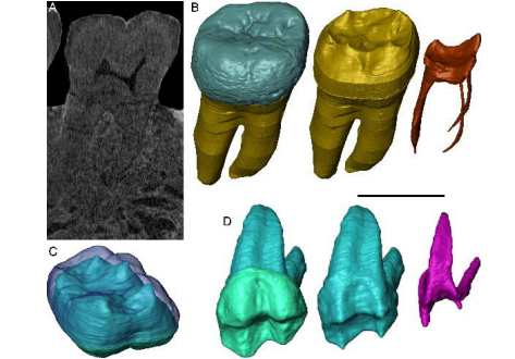
Exploring hominin and non-hominin primate dental fossil remains with neutron microtomography
Fossil dental remains are an archive of unique information for paleobiological studies. Computed microtomography based on Xray microfocus sources (X-µCT) and Synchrotron Radiation (SR-µCT) allow subtle quantification at the micron and sub-micron scale of the meso- and microstructural signature imprinted in the mineralized tissues, such as enamel and dentine, through highresolution “virtual histology”. Nonetheless, depending on the degree of alterations undergone during fossiliza... Read more
Clément Zanolli, Laboratory AMIS, UMR 5288, University of Toulouse III - Paul Sabatier, France, and al.
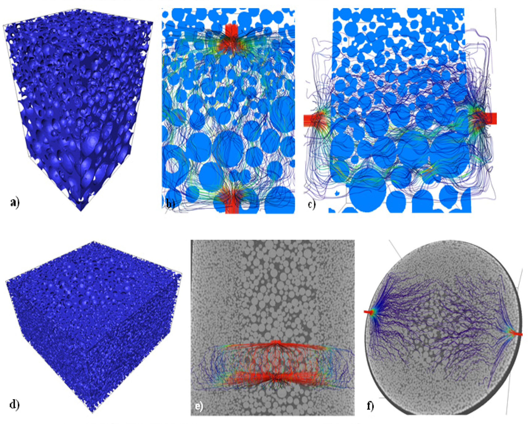
Nowadays, industrial processes demand materials with specific properties and localized microstructures to improve material performance. To satisfy particular needs, the development of materials with changing mechanical properties and/or microstructures along a preferential direction has been developed. These are called Functional Graded Materials (FGMs). Among these materials, a variation on the porosity along the part is very useful for different industrial applications, such as microfiltrat... Read more
Jorge Sergio Téllez-Martínez, Luis Olmos, Víctor Manuel Solorio-García, Héctor Javier Vergara-Hernández, Jorge Chávez, Dante Arteaga
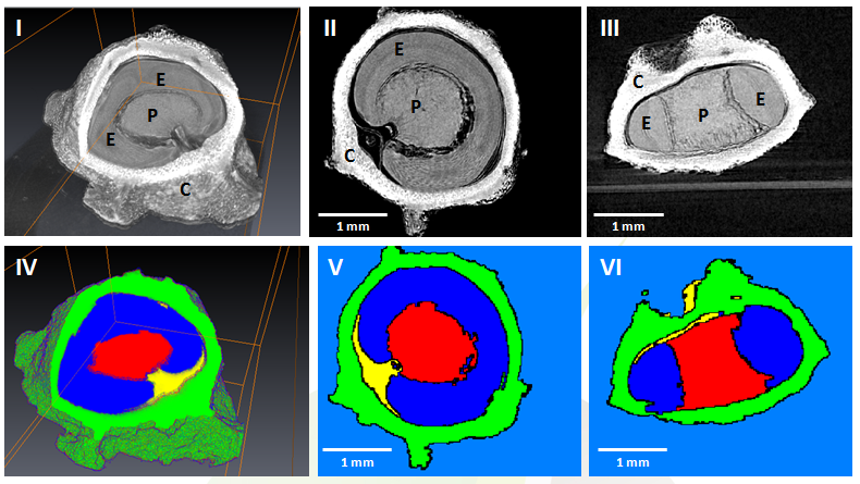
GEVES uses Avizo software to study and control plant seeds
At GEVES, Microtomography (micro-CT), which is a technique using X-rays to investigate internal anatomy and morphology of organisms without destruction, is applied to plant seed quality control and phenotyping. Several applications have been developed using micro-CT coupled with Avizo software development, such as for example in the Measurements of internal and external sugar beet seed structures for seed phenotyping
Read more
Ghassen Trigui, Laurence Le Corre, Dominique Honoré and Karima Boudehri-Giresse - 2D/3D Imaging - R&D team, GEVES
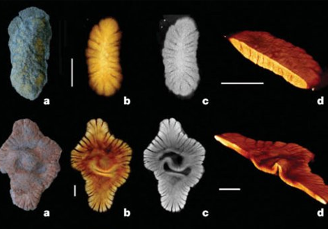
2 BILLION years old fossils appear to represent a first experiment in megascopic multicellularity
The Paleoproterozoic Era witnessed crucial steps in the evolution of Earth’s surface environments following the first appreciable rise of free atmospheric oxygen concentrations ∼2.3 to 2.1 Ga ago, and concomitant shallow ocean oxygenation. Combined microtomography, geochemistry, and sedimentary analysis suggest a biota fossilized during early diagenesis. The emergence of this biota follows a rise in atmospheric oxygen, which is consistent with the idea that surface oxygenation allowe... Read more
Abderrazak El Albani, Laboratoire HYDRASA, UMR 6269 CNRS-INSU, Université de Poitiers, France
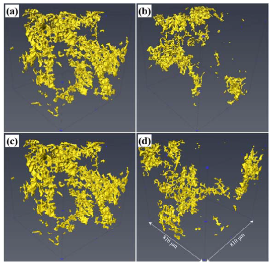
The effect of ultrasonic melting processing on three-dimensional architecture of intermetallic phases and pores in two multicomponent cast Al-5.0Cu 0.6Mn-0.5 Fe alloys is characterized using conventional microscopy and synchrotron X-ray microtomography. (…) The results show that ultrasonic melt processing (USP) significantly reduce the volume fraction, grain size, interconnectivity, and equivalent diameter of the intermetallic phases in both alloys. The volume fraction of pores in both ... Read more
Yuliang Zhao, Dongfu Song, Bo Lin, Chun Zhang, Donghai Zheng, Zhi Wang, Weiwen Zhang
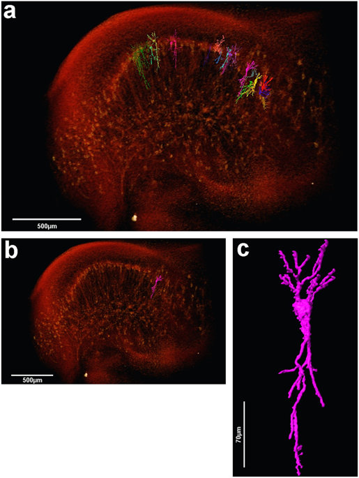
The assessment of neuronal number, spatial organization and connectivity is fundamental for a complete understanding of brain function. However, the evaluation of the three-dimensional (3D) brain cytoarchitecture at cellular resolution persists as a great challenge in the field of neuroscience. In this context, X-ray microtomography has shown to be a valuable non-destructive tool for imaging a broad range of samples, from dense materials to soft biological specimens, arisen as a new method fo... Read more
Matheus de Castro Fonseca, Bruno Henrique Silva Araujo, Carlos Sato Baraldi Dias, Nathaly Lopes Archilha, Dionísio Pedro Amorim Neto, Esper Cavalheiro, Harry Westfahl Jr, Antônio José Roque da Silva, Kleber Gomes Franchini
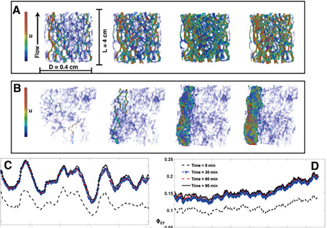
We have experimentally investigated the impact of heterogeneity on the dissolution of two limestones, characterised by distinct degrees of flow heterogeneity at both the pore and core scales. The two rocks were reacted with reservoir-condition CO2-saturated brine at both scales and scanned dynamically during dissolution. First, 1 cm long 4 mm... Read more
Menke H.P; Reynolds C.A.; Andrew M.G.; Pereira Nunes J.P.; Bijeljic B; Blunt M.J.

Metal additive manufacturing techniques such as the powder-bed systems are developing as a novel method for producing complex components.
This study uses synchrotron-based X-ray microtomography to investigate porosity in electron beam melted Ti-6Al-4V in the as-built and post-processed state for two different powders. The presence of gas porosity in the starting powder was shown to correlate to porosity in the as-built components. This porosity was observed to shrink after a hot isosta... Read more
Ross Cunningham, Andrea Nicolas, John Madsen, Eric Fodran, Elias Anagnostou, Michael D. Sangid & Anthony D. Rollett
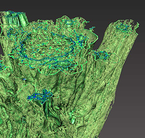
Unilever uses Avizo software to visualize and understand food and detergent structures
Food and detergent products are composed of complex micro structures. With modern microscopic techniques we can make them visible. The microstructure greatly affects macroscopic properties such as appearance, taste, mouth feel and solubility. Making these structures visible and quantifying them is essential to the development of products with optimal product properties. A broad range of imaging techniques is used to visualize microstructure elements at different length scales. For example, X-... Read more
Gerard van Dalen, Unilever R&D Vlaardingen (The Netherlands)
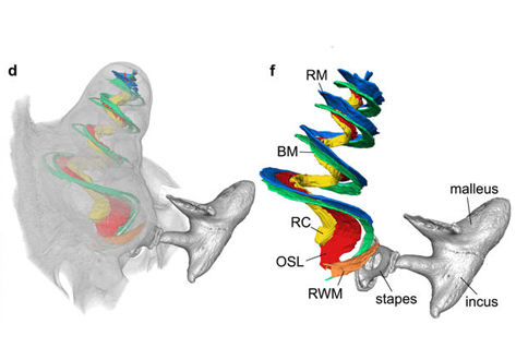
Propagation-based phase-contrast x-ray tomography of cochlea using a compact synchrotron source
We demonstrate that phase retrieval and tomographic imaging at the organ level of small animals can be advantageously carried out using the monochromatic radiation emitted by a compact x-ray light source, without further optical elements apart from source and detector. This approach allows to carry out microtomography experiments which – due to the large performance gap with respect to conventional laboratory instruments – so far were usually limited to synchrotron sources. We dem... Read more
Mareike Töpperwien, Regine Gradl, Daniel Keppeler, Malte Vassholz, Alexander Meyer, Roland Hessler, Klaus Achterhold, Bernhard Gleich, Martin Dierolf, Franz Pfeiffer, Tobias Moser & Tim Salditt