Welcome to the Amira-Avizo Software Use Case Gallery
Below you will find a collection of use cases of our 3D data visualization and analysis software. These use cases include scientific publications, articles, papers, posters, presentations or even videos that show how Amira-Avizo Software is used to address various scientific and industrial research topics.
Use the Domain selector to filter by main application area, and use the Search box to enter keywords related to specific topics you are interested in.
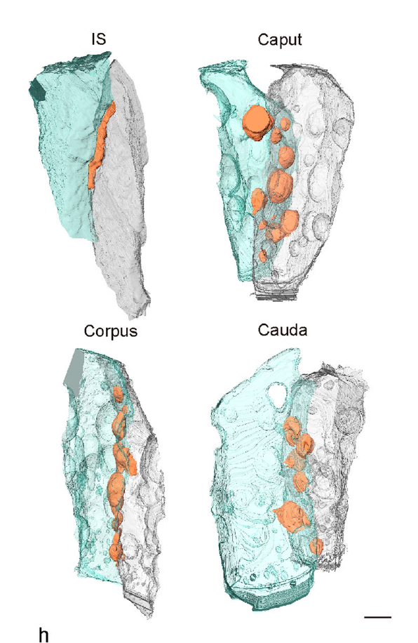
Mammalian epididymal epithelial cells are crucial for sperm maturation. Historically,
vacuole-like ultrastructures in epididymal epithelial cells were
observed via transmission electron microscopy but were undefined. Here, we
utilize volume electron microscopy (vEM) to generate 3D reconstructions of
epididymal epithelial cells and identify these vacuoles as intercellular organelle
reservoirs (IORs) in the lateral intercellular space (LIS), which contains
pr... Read more
Xia Li, Feng Qiao, Jiansheng Guo, Ting Jiang, Huifang Lou, Huixia Li, Gangcai Xie, Hangjun Wu, Weizhen Wang, Ruoyu Pei, Sha Liu, Mei Ye, Jin Li, Shiqin Huang, Mengya Zhang, Chaoye Ma, Yiwen Huang, Shushu Xu, Xiaofeng Li, Xiao Sun, Jun Yu, Kin Lam Fok, Shumin Duan & Hao Chen
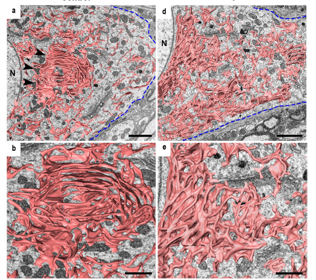
The endoplasmic reticulum (ER) extends throughout a cell and plays a critical role in maintaining cellular homeostasis.
Changes in ER shape could provide a clue to explore the mechanisms that underlie the fate determination of neurons after
axon injury because the ER drastically changes its morphology under neuronal stress to maintain cellular homeostasis and
recover from damage. Because of their tiny structures and richness in the soma, the detailed morphology of the ER and... Read more
Mahmoud Elgendy,Hiromi Tamada, Takaya Taira, Yuma Iio, Akinobu Kawamura, Ayusa Kunogi, Yuka Mizutani, Hiroshi Kiyama
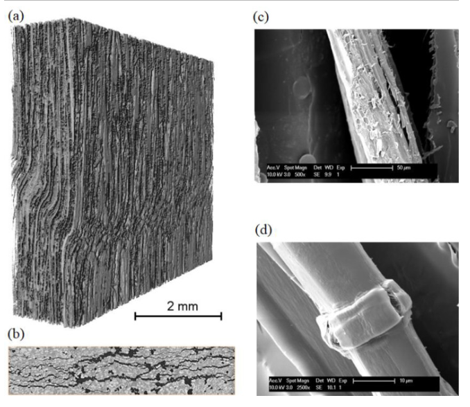
In this study, the researchers investigated the compressive failure mechanisms in flax fiber composites, a promising eco-friendly alternative to synthetic composite materials, through both numerical simulations and experimental analysis. They examined the reasons behind the low compressive strength in comparison to tensile strength, focusing on the compressive-to-tensile strength ratio. A novel thermodynamically consistent continuum damage micromechanics model was introduced to capture the ev... Read more
Vedad Tojaga, Alexandros Prapavesis, Jonas Faleskog, T. Christian Gasser, Aart W. van Vuure, Sören Östlund
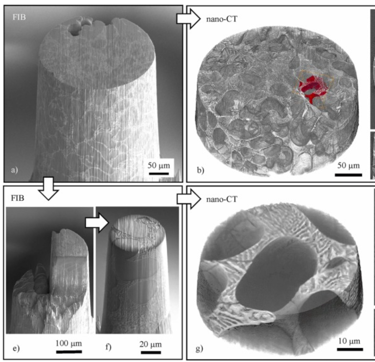
Three-dimensional imaging of microstructural evolution in SEM-based nano-CT
Scanning electron microscopy (SEM) is a powerful and versatile technique for materials characterization and present in many laboratories. The integration of an X-ray target holder and detector allows expanding the modalities of SEM by X-ray imaging. These little hardware adaptations enable radiography ... Read more
Jonas Fell, Christoph Pauly, Michael Maisl, Simon Zabler, Frank Mücklich, Hans-Georg Herrmann
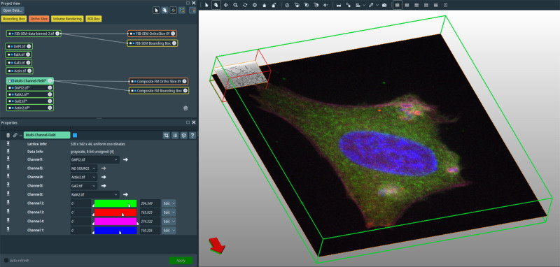
In recent years new methodologies and workflow pipelines for acquiring correlated fluorescence microscopy and volume electron microscopy datasets have been extensively described and made accessible to users of different levels. Post-acquisition image processing, and particularly correlation of the optical and electron data in a single integrated three-dimensional framework can be key for extracting valuable information, especially when imaging large sample volumes such as whole cells or tissu... Read more
Allon Weiner
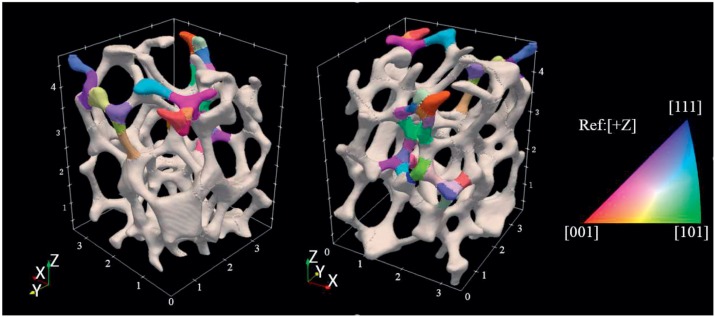
The complex mechanical response of open-cell foams depends strongly on the hierarchy of length scales inherent in them, from engineering-part scale to the ligament scale through the grain scale down to the crystal-lattice scale. A first step toward understanding and predicting the coordinated mechanical response across le... Read more
Jayden C. Plumb, Jonathan F. Lind, Joseph C. Tucker, Ron Kelley, Ashley D. Spear
Multi-modal plasma focused ion beam serial section tomography of an organic paint coating
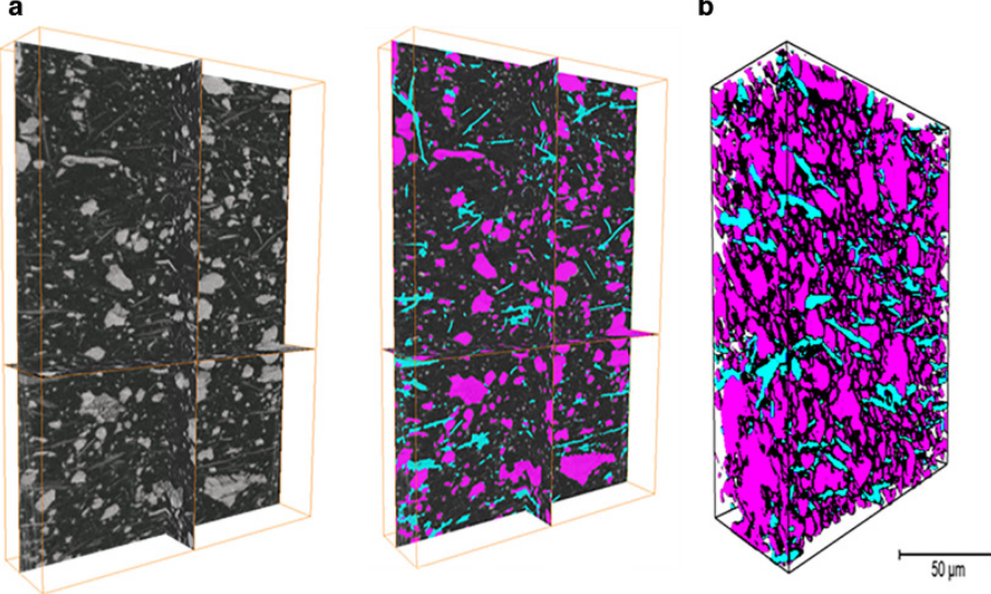
Pigment distributions have a critical role in the corrosion protection properties of organic paint coatings, but they are difficult to image in 3D over statistically significant volumes and at sufficiently high spatial resolutions required for detailed analysis. Here we report, for the first time, large volume analytical serial sectioning tomography of an organic composite coating using a xenon Plasma Focused Ion Beam (PFIB) combined with secondary electron imaging, energy dispersive X-ray (E... Read more
Zhong Xiangli, M. Grace Burke, Philip J. Withers, Zhang Xun, Zhou Xiaorong, Timothy L. Burnett, Liu Yanwen, Stuart B. Lyon, Simon R.Gibbon
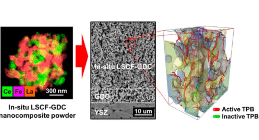
Improving microstructural quantification in FIB/SEM nanotomography
Advanced nanotomographic analysis is still far from routine, and a number of challenges remain in data acquisition and post-processing. In this work, we present a number of techniques to improve the quality of the acquired data, together with easy-to-implement methods to obtain “advanced” microstructural quantifications. The techniques are applied to a solid oxide fuel cell cathode of interest to the electrochemistry community, but the methodologies are easily adaptable to a wide range of... Read more
Joshua A.Taillon, Christopher Pellegrinelli, Yi-Lin Huang, Eric D.Wachsman, Lourdes G. Salamanca-Riba
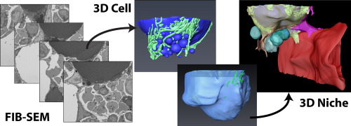
Characterization of the bone marrow adipocyte niche with three-dimensional electron microscopy
Unlike white and brown adipose tissues, the bone marrow adipocyte (BMA) exists in a microenvironment containing unique populations of hematopoietic and skeletal cells.
To study this microenvironment at the subcellular level, we performed a three-dimensional analysis of the ultrastructure of the BMA niche with focused ion beam scanning electron microscopy (FIB-SEM). This revealed that BMAs display hallmarks of metabolically active cells including polarized lipid deposits, a dense mitoch... Read more
Hero Robles, SungJae Park, Matthew S. Joens, James A.J. Fitzpatrick, Clarissa S. Craft, Erica L. Scheller
Composite cathodes comprising nanoscale
powders are expected to impart with high specific surface
area and triple phase boundary (TPB) density, which will lead
to better performance.
However, uniformly mixing nanosized heterophase powders remains a challenge due to their high surface energy and thus ease with which they agglomerate into their individual phases during the mixing and sintering
processes. In this study, we successfully synthesized La0.6Sr0.4Co0.2Fe... Read more
Dong Woo Joh, Areum Cha, Jeong Hwa Park, Kyeong Joon Kim, Kyung Taek Bae, Doyeub Kim, Young Ki Choi, Hyegsoon An, Ji Su Shin, Kyung Joong Yoon, and Kang Taek Lee
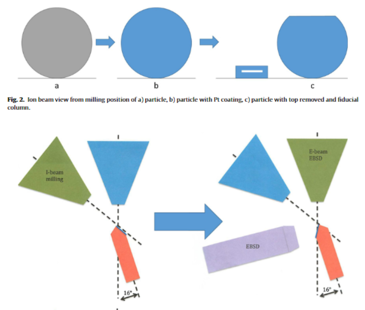
Automated 3D EBSD for metallic powders
Metallic powders are commonly used in additive manufacturing processes. While their post-process consolidated properties are widely studied, there is little research on the properties of the powders prior to consolidation. Understanding the powder characteristics before use in additive manufacturing processes could lead to fine-tuning properties of additively manufactured materials. The three-dimensional grain structure of metals can be useful in predicting their properties and ... Read more
Caitlin Walde, Roger Ristau, Danielle Cote
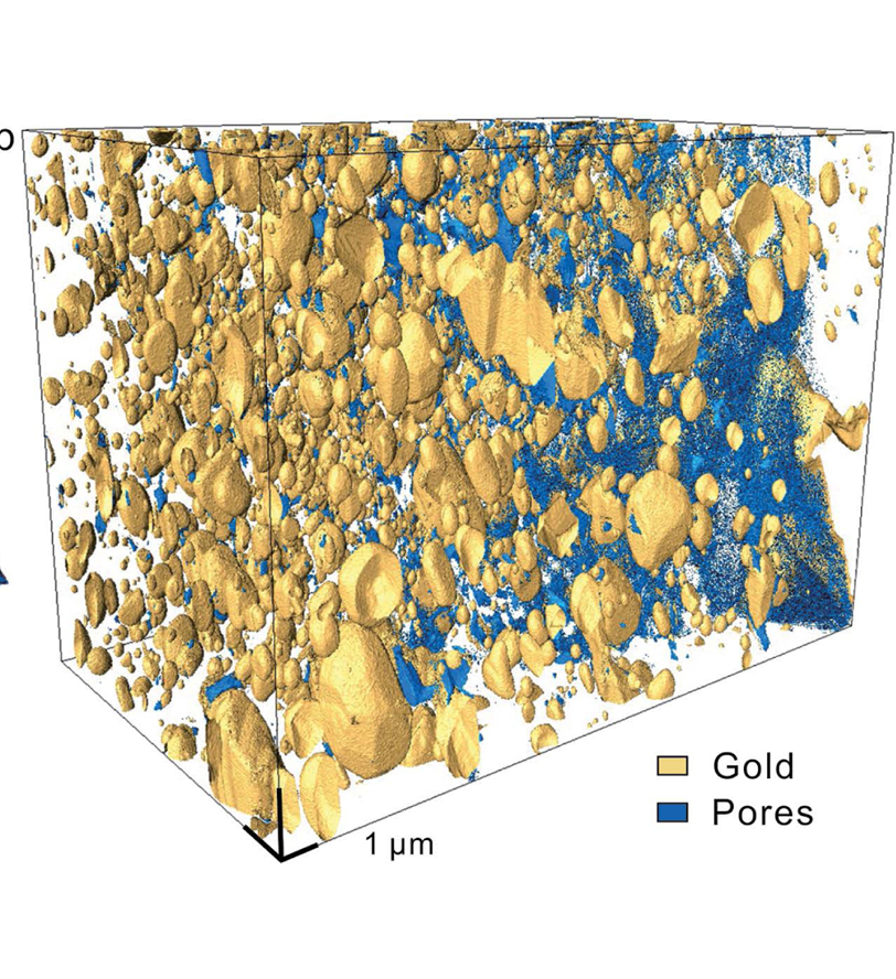
Recent studies have identified gold nanoparticles in ores in a range of deposit types, but little is known about their formation processes. In this contribution, gold-bearing magnetite from the well-documented, world-class Beiya Au deposit, China, was investigated in terms of microstructure and crystallography at the nanoscale. We present the first three-dimensional (3D) focused ion beam/scanning electron microscopy (FIB/SEM) tomography of the distribution of gold nanoparticles in nanopores i... Read more
Haoyang Zhou, Richard Wirth, Sarah A. Gleeson, Anja Schreiber, Sathish Mayanna
Cryo-STEM mapping of solid–liquid interfaces and dendrites in lithium-metal batteries
Solid–liquid interfaces are important in a range of chemical, physical and biological processes but are often not fully understood owing to the lack of high-resolution characterization methods that are compatible with both solid and liquid components. For example, the related processes of dendritic deposition of lithium metal and the formation of solid–electrolyte interphase layers are known to be key determinants of battery safety and performance in high-energy-density lithium-metal bat... Read more
Michael J. Zachman, Zhengyuan Tu, Snehashis Choudhury, Lynden A. Archer & Lena F. Kourkoutis
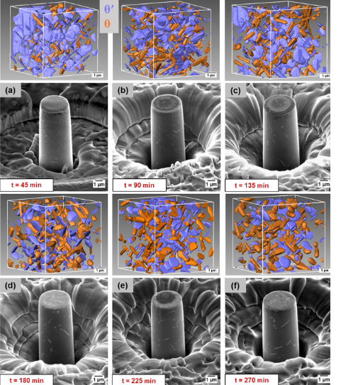
A unique approach to correlating an evolving 3D microstructure in an Al-Cu alloyand its micro-scale mechanical properties has been introduced. Using these nanoscale three-dimensional microstructures derived from Transmission X-rayMicroscopy (TXM), individual contributions from different strengthening mechanisms were quantified. The spatial distribution and morphology of the individual θ′ and θ phases were seen to play an important role in influencing dislocation storage. Uniaxi... Read more
C. Shashank Kaira, Christopher Kantzos, Jason J. Williams, Vincent De Andrade, Francesco De Carlo, Nikhilesh Chawlaa
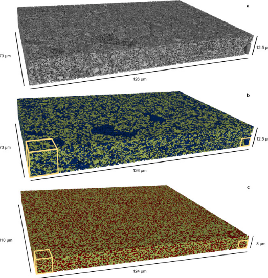
Mesoscale characterization of local property distributions in heterogeneous electrodes
The performance of electrochemical devices depends on the three-dimensional (3D) distributions of microstructural features in their electrodes. Several mature methods exist to characterize 3D microstructures over the microscale (tens of microns), which are useful in understanding homogeneous electrodes. However, methods that capture mesoscale (hundreds of microns) volumes at appropriate resolution (tens of nm) are lacking, though they are needed to understand more common, less ideal electrode... Read more
Tim Hsu, William K. Epting, Rubayyat Mahbub, Noel T. Nuhfer, Sudip Bhattachary, Yinkai Lei, Herbert M. Miller, Paul R. Ohodnicki, Kirk R. Gerdes, Harry W. Abernathy, Gregory A. Hackett, Anthony D. Rollett, Marc De Graef, Shawn Litster, Paul A. Salvador
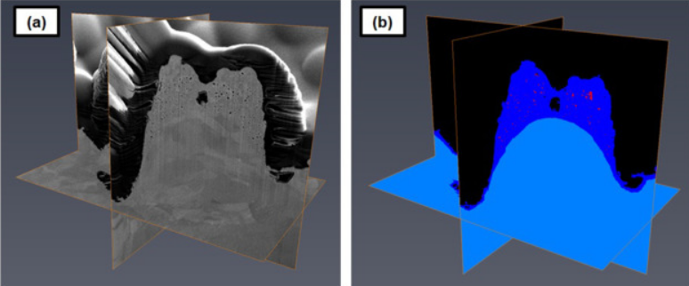
Laser processing of metal surfaces by ultrafast Read more
Edwin Peng, Alexander Roth, Craig A. Zuhlke, Soodabeh Azadehranjbar, Dennis R. Alexander, George Gogos, Jeffrey E. Shield
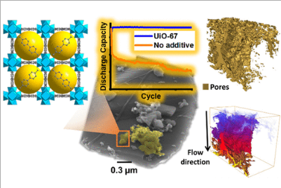
Porous Metal–Organic Frameworks for Enhanced Performance Silicon Anodes in Lithium-Ion Batteries
Maintaining the physical integrity of electrode microstructures in Li-ion batteries is critical to significantly extend their cycle life. This is especially important for high-capacity anode materials such as silicon, whose operational volume expansion exerts huge internal stress within the anode, resulting in electrode destruction and capacity fade. In this study, we demonstrate that by incorporating metal–organic frameworks (MOFs) with carboxylate organic linkers into Si-based anodes, a s... Read more
Romeo Malik, Melanie. J. Loveridge, Luke J. Williams, Qianye Huang, Geoff West, Paul R. Shearing, Rohit Bhagat, Richard I. Walton
Microstructural analysis of TRISO particles using multi-scale X-ray computed tomography
TRISO particles, a composite nuclear fuel built up by ceramic and graphitic layers, have outstanding high temperature resistance. TRISO fuel is the key technology for High Temperature Reactors (HTRs) and the Generation IV Very High Temperature Reactor (VHTR) variant.
TRISO offers unparalleled containment of fission products and is extremely robust during accident conditions. An understanding of the thermal performance and mechanical properties of TRISO fuel requires a detailed knowledg... Read more
T. Lowe, R.S. Bradley, S. Yue, K. Barii, J. Gelb, N. Rohbeck, J. Turner, P.J. Withers
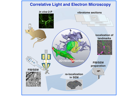
Label-free 3D-CLEM using endogenous tissue landmarks
We demonstrate feasibility of the workflow by combining in vivo 2-photon microscopy and focused ion beam scanning electron microscopy (FIB/SEM) to dissect the role of astrocytic coverage in the persistence of dendritic spines.
Emerging 3D correlative light and electron microscopy (CLEM) approaches enable studying neuronal structure-function relations at unprecedented depth and precision. However, established protocols for the correlation of light and electron micrographs rely ... Read more
Manja Luckner,Steffen Burgold, Severin Filser, Maximilian Scheungrab, Yilmaz Niyaz, Eric Hummel, Gerhard Wanner, Jochen Herms
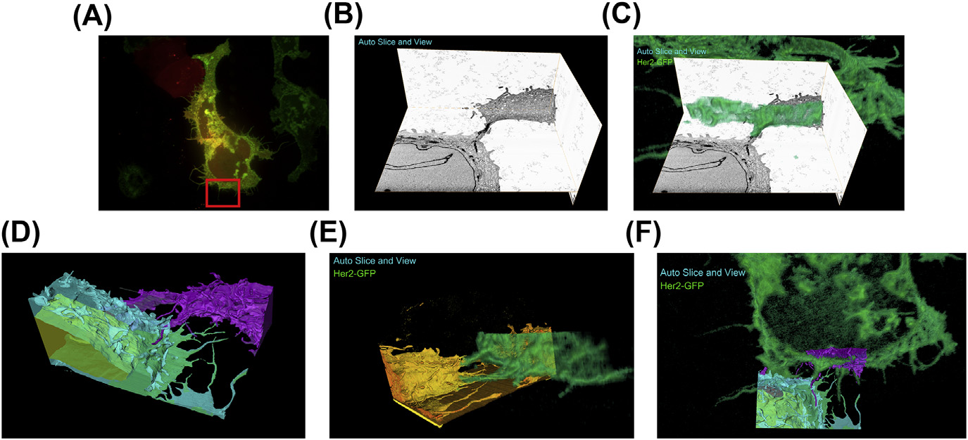
A fully integrated, three-dimensional fluorescence to electron microscopy correlative workflow
While fluorescence microscopy provides tools for highly specific labeling and sensitive detection, its resolution limit and lack of general contrast has hindered studies of cellular structure and protein localization. Recent advances in correlative light and electron microscopy (CLEM), including the fully integrated CLEM workflow instrument, the Thermo Scientific CorrSight with MAPS, have allowed for a more reliable, reproducible, and quicker approach to correlate three-dimensional time-lapse... Read more
Claudia S. Lopez, Cedric Bouchet-Marquis, Christopher P. Arthur, Jessica L. Riesterer, Gregor Heiss, Guillaume Thibault, Lee Pullan, Sunjong Kwon, Joe W. Gray

Synergistic role of nucleotides and lipids for the self-assembly of Shs1 septin oligomers
Amira capacities for membranes and filaments segmentation in cryo-TEM images are featured on the front cover of Biochemical Journal, July 2020.
Budding yeast septins are essential for cell division and polarity. (…) [The authors] have dissected, here, for the first time, the behavior of the Shs1 protomer bound to membranes at nanometer resolution, in complex with the other septins. Using electron microscopy, [the authors] have shown that on membranes, Shs1 protomers self-assembl... Read more
Cyntia Taveneau, Rémi Blanc, Gerard Pehau-Arnaudet, Aurélie Cicco, Aurélie Bertin