Welcome to the Amira-Avizo Software Use Case Gallery
Below you will find a collection of use cases of our 3D data visualization and analysis software. These use cases include scientific publications, articles, papers, posters, presentations or even videos that show how Amira-Avizo Software is used to address various scientific and industrial research topics.
Use the Domain selector to filter by main application area, and use the Search box to enter keywords related to specific topics you are interested in.
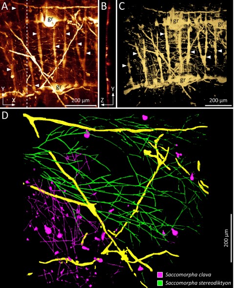
Microscopic organisms that penetrate calcareous structures by actively dissolving the carbonate matrix, namely microendoliths, have an important influence on the breakdown of marine carbonates.
Microscopic organisms that penetrate calcareous structures by actively dissolving the carbonate matrix, namely microendoliths, have an important influence on the breakdown of marine carbonates. The study of these microorganisms and the bioerosion traces they produce is crucial for understanding ... Read more
Philipp-Konrad Schätzle, Max Wisshak, Andreas Bick, André Freiwald, Alexander Kieneke

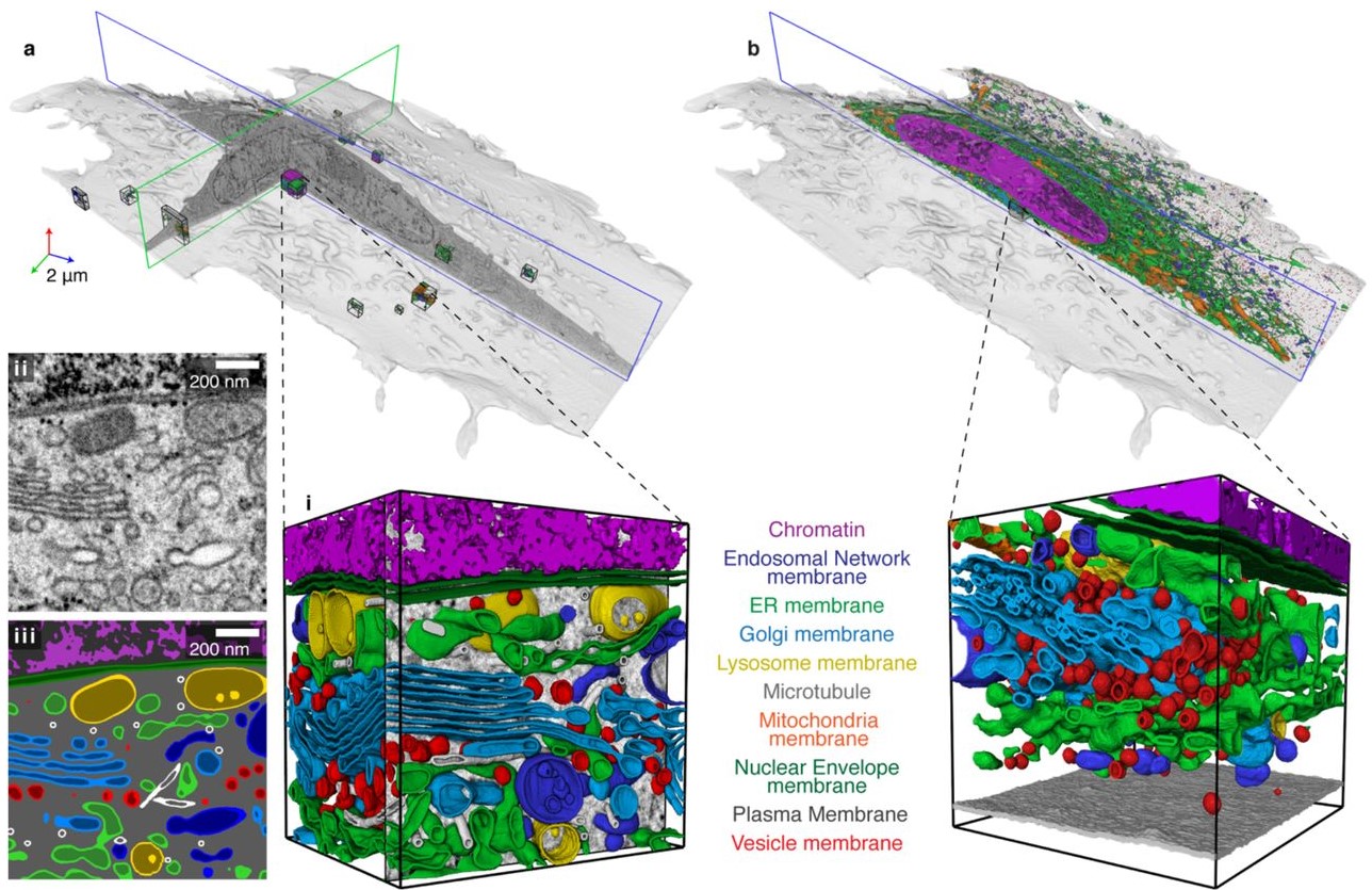
Protocols for Generating Surfaces and Measuring 3D Organelle Morphology Using Amira
High-resolution 3D images of organelles are of paramount importance in cellular biology. Although light microscopy and transmission electron microscopy (TEM) have provided the standard for imaging cellular structures, they cannot provide 3D images.
However, recent technological advances such as serial block-face scanning electron microscopy (SBF-SEM) and focused ion beam scanning electron microscopy (FIB-SEM) provide the tools to create 3D images for the ultrastructural analysis of org... Read more
Edgar Garza-Lopez, Zer Vue, Prasanna Katti, Kit Neikirk, Michelle Biete, Jacob Lam, Heather K. Beasley, Andrea G. Marshall, Taylor A. Rodman, Trace A. Christensen, Jeffrey L. Salisbury, Larry Vang, Margaret Mungai, Salma Ash Shareef, Sandra A. Murray, Jianqiang Shao, Jennifer Streeter, Brian Glancy, Renata O. Pereira1, E. Dale Abel, and Antentor Hinton, Jr.
Automatic whole cell organelle segmentation in volumetric electron microscopy
Cells contain hundreds of different organelle and macromolecular assemblies intricately organized relative to each other to meet any cellular demands. Obtaining a complete understanding of their organization is challenging and requires nanometer-level, three-dimensional reconstruction of whole cells. Even then, the immense size of datasets and large number of structures to be characterized requires generalizable, automatic methods.
To meet this challenge, we developed an analy... Read more
Larissa Heinrich, Davis Bennett, David Ackerman, Woohyun Park, John Bogovic, View ORCID ProfileNils Eckstein, Alyson Petruncio, Jody Clements, C. Shan Xu, Jan Funke, Wyatt Korff, Harald F. Hess, Jennifer Lippincott-Schwartz, Stephan Saalfeld, Aubrey V. Weigel, COSEM Project Team
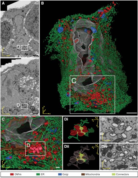
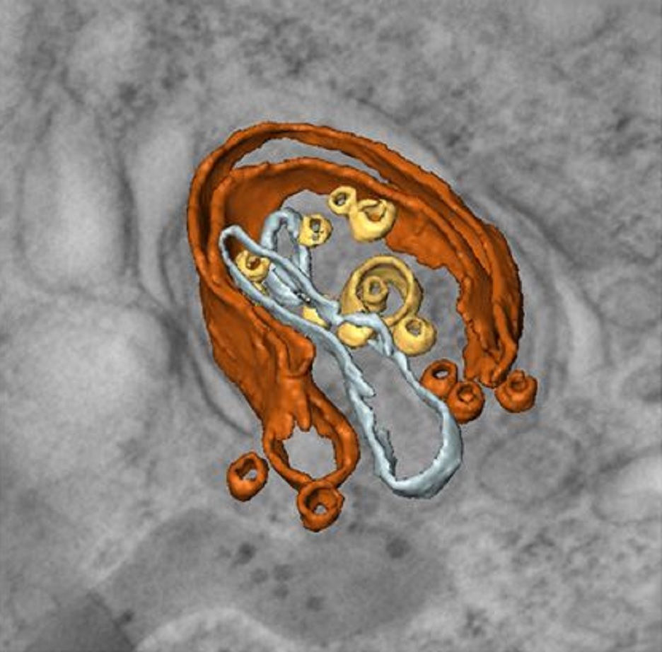
Integrative Imaging Reveals SARS-CoV-2-Induced Reshaping of Subcellular Morphologies
Cortese et al. use integrative imaging techniques to generate a publicly available repository of morphological alterations induced by SARS-CoV-2 in lung cells. Accumulation of ER-derived double-membrane vesicles, the viral replication organelle, occurs concomitantly with cytoskeleton remodeling and Golgi fragmentation. Pharmacological alteration of cytoskeleton dynamics restricts viral replication and spread.
Pathogenesis induced by SARS-CoV-2 is thought to result from both an inflamma... Read more
Mirko Cortese, Ji-Young Lee, Berati Cerikan, ..., Laurent Chatel-Chaix, Yannick Schwab, Ralf Bartenschlager
Macroautophagy is morphologically characterized by autophagosome formation. Autophagosomes are double-membraned vesicles that sequester cytoplasmic components for further degradation in the lysosome. Basal autophagy is paramount for intracellular quality control in post-mitotic cells but, surprisingly, the number of autophagosomes in post-mitotic neurons is very low, suggesting that alternative degradative structures could exist in neurons…
Read more
Maria Rosario Fernandez-Fernandez, Desire Ruiz-Garcia, Eva Martin-Solana, Francisco Javier Chichon, Jose L. Carrascosa, Jose-Jesus Fernandez
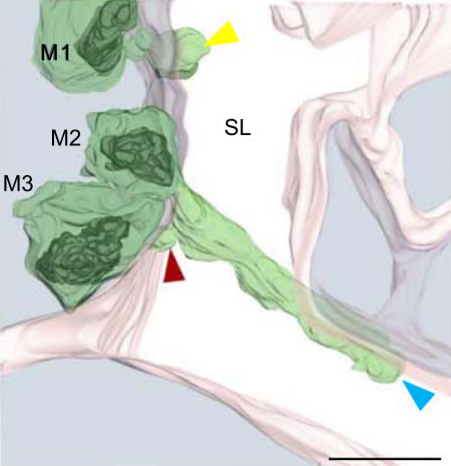
Influenza A matrix protein M1 is sufficient to induce lipid membrane deformation
The matrix protein M1 of the Influenza A virus is considered to mediate viral assembly and budding at the plasma membrane (PM) of infected cells. In order for a new viral particle to form, the PM lipid bilayer has to bend into a vesicle towards the extracellular side. Studies in cellular models have proposed that different viral proteins might be responsible for inducing membrane curvature in this context (including M1), but a clear consensus has not been reached. In this study, we use a comb... Read more
Ismail Dahmani, Kai Ludwig, Salvatore Chiantia
Multiple membrane extrusion sites drive megakaryocyte migration into bone marrow blood vessels
Platelets, cells central to hemostasis and thrombosis, are formed from parent cell megakaryocytes. Although the process is highly efficient in vivo, our ability to generate them in vitro is still remarkably inefficient. We proposed that greater understanding of the process in vivo is needed and used an imaging approach, intravital correlative light electron microscopy, to visualize platelet generation in bone marrow in the living mouse. In contrast to current understanding, we found that most... Read more
Edward Brown, Leo M Carlin, Claus Nerlov, Cristina Lo Celso, Alastair W Poole
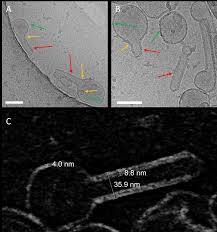
Membrane architecture of pulmonary lamellar bodies revealed by post-correlation on-lamella cryo-CLEM
Lamellar bodies (LBs) are surfactant rich organelles in alveolar type 2 cells. LBs disassemble into a lipid-protein network that reduces surface tension and facilitates gas exchange at the air-water interface in the alveolar cavity. Current knowledge of LB architecture is predominantly based on electron microscopy studies using disruptive sample preparation methods. We established a post-correlation on-lamella cryo-correlative light and electron microscopy approach for cryo-FIB milled lung ce... Read more
Steffen Klein, Benedikt H. Wimmer, Sophie L. Winter, Androniki Kolovou, Vibor Laketa, Petr Chlanda
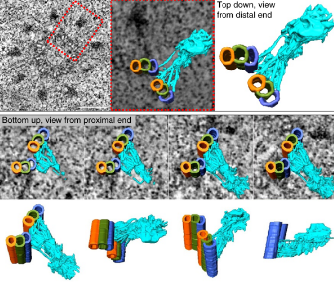
Centrioles are vital cellular structures that form centrosomes and cilia. The formation and function of cilia depends on a set of centriole’s distal appendages. In this study, we use correlative super resolution and electron microscopy to precisely determine where distal appendage proteins localize in relation to the centriole microtubules and appendage electron densities. Here we characterize a novel distal appendage protein ANKRD26 and detail, in high resolution, the initial steps of dist... Read more
Mathew Bowler, Dong Kong, Shufeng Sun, Rashmi Nanjundappa, Lauren Evans, Veronica Farmer, Andrew Holland, Moe R. Mahjoub, Haixin Sui & Jadranka Loncarek

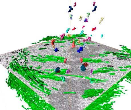
Soluble tubulin is locally enriched at mitotic centrosomes in C. elegans
During mitosis, the centrosome expands its capacity to nucleate microtubules. Understanding the mechanisms of centrosomal microtubule nucleation is, however, constrained by a lack of knowledge of the amount of soluble and polymer tubulin at mitotic centrosomes. Here we combined light microscopy and serial-section electron tomography to measure the amount of dimer and polymer at mitotic centrosomes in early C. elegans embryos. We show that a C. elegans one-cell stage centrosome at metaphase co... Read more
Johannes Baumgart, Marcel Kirchner, Stefanie Redemann, Jeffrey Woodruff, Jean-Marc Verbavatz, Frank Julicher, Anthony Hyman, Thomas Mueller-Reichert, Jan Brugues
Serial block face scanning electron microscopy (SBF-SEM) is a powerful method to analyze cells in 3D. Here, working at the resolution limit of the method, we describe a correlative light–SBF-SEM workflow to resolve microtubules of the mitotic spindle in human cells. We present four examples of uses for this workflow that are not practical by light microscopy and/or transmission electron microscopy. First, distinguishing closely associated microtubules within K-fibers; second, resolving brid... Read more
Faye M. Nixon, Thomas R. Honnor, Nicholas I. Clarke, Georgina P. Starling, Alison J. Beckett, Adam M. Johansen, Julia A. Brettschneider, Ian A. Prior, Stephen J. Royle
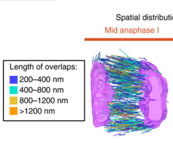
Chromosome segregation occurs by microtubule pushing in oocytes
During cell division, spindle microtubules ensure an equal repartition of chromosomes between the two daughter cells. While the kinetochore-dependent mechanisms that drive mitotic chromosome segregation are well understood, in oocytes of most species atypical spindles assembled in absence of centrosomes entail poorly understood mechanisms of chromosome segregation. In particular, the structure(s) responsible for force generation during meiotic chromosome separation in oocytes is unclear. Usin... Read more
Kimberley Laband, Rémi Le Borgne, Frances Edwards, Marine Stefanutti, Julie C. Canman, Jean-Marc Verbavatz, Julien Dumont
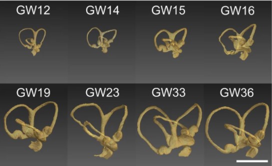
Progressive transformation of the otic placode into the functional inner ear during gestational development in humans leads to the acquisition of hearing perception via the cochlea and balance and spatial orientation via the vestibular organ.
Using a correlative approach involving micro-computerized tomography (micro-CT), transmission electron microscopy and histological techniques we were able to examine both the morphological and cellular changes associated with human inner ear devel... Read more
Lejo Johnson Chacko, David Wertjanz, Consolato Sergi, Jozsef Dudas, Natalie Fischer, Theresa Eberharter, Romed Hoermann, Rudolf Glueckert, Helga Fritsch, Helge Rask-Andersen, Anneliese Schrott-Fischer & Stephan Handschuh
In biomedical research, a huge variety of different techniques is currently available for the structural examination of small specimens, including conventional light microscopy (LM), transmission electron microscopy (TEM), confocal laser scanning microscopy (CLSM), microscopic X-ray computed tomography (microCT), and many others. Since every imaging method is physically limited by certain parameters, a correlative use of complementary methods often yields a significant broader range of inform... Read more
Stephan Handschuh, Natalie Baeumler, Thomas Schwaha & Bernhard Ruthensteiner
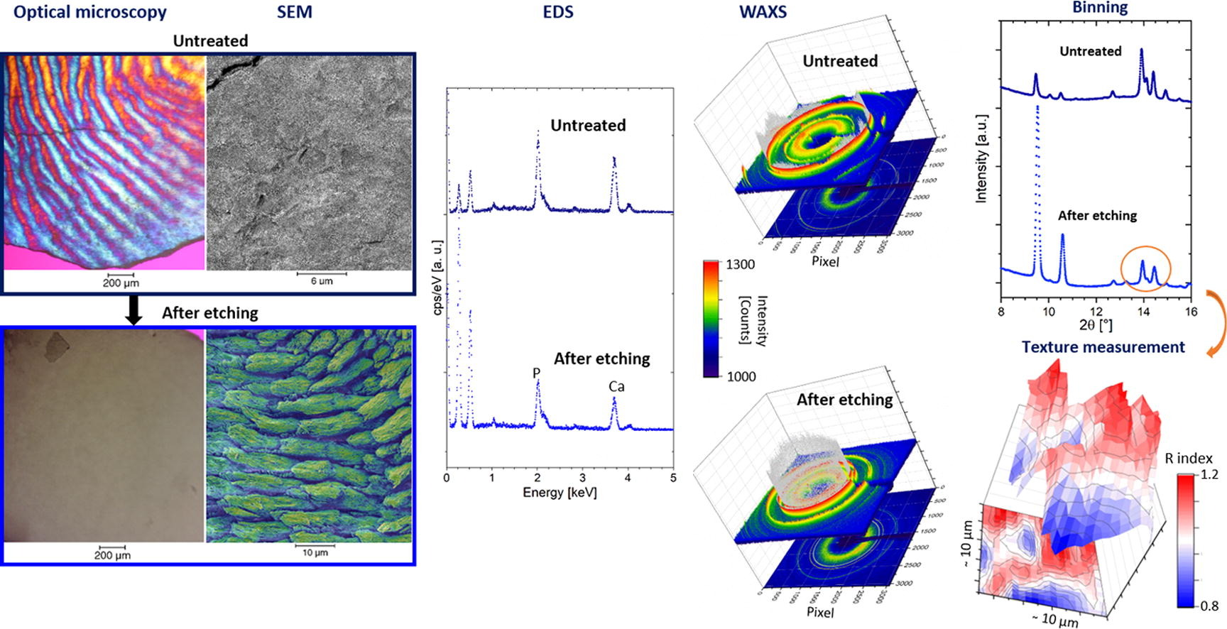
Enamel caries is a highly prevalent worldwide disease that involves the demineralisation of the outer tooth structure. In this study, we report the analysis of artificially demineralised human enamel sections (‘slices’) etched using lactic acid (2% v/v) in comparison with healthy enamel using correlative techniques of optical and electron microscopy, as well as scanning diffraction. Demineralisation of the enamel was characterised at the micron to sub-micron scale. The structure of the he... Read more
Cyril Besnard, Robert A. Harper, Thomas E. J. Moxham, Jonathan D. James, Malte Storm, Enrico Salvati, Gabriel Landini, Richard M. Shelton, Alexander M.Korsunsky
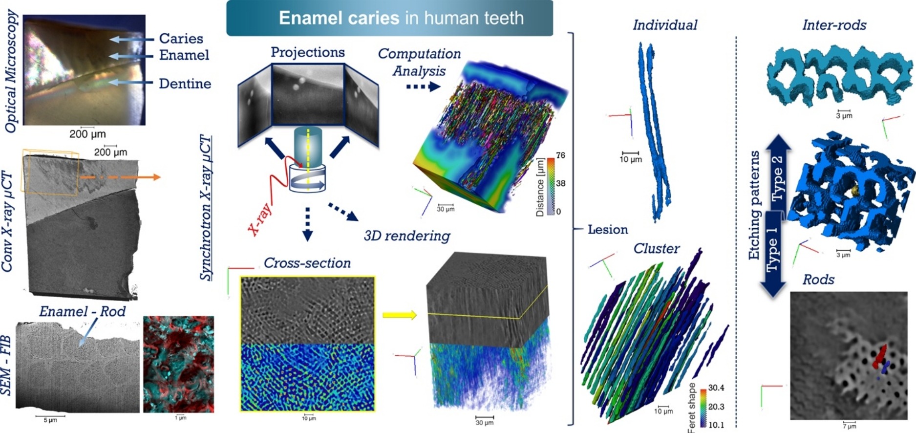
Unprecedented combination of resolution, field of view and contrast for the analysis human enamel carious lesions was achieved. Synchrotron X-ray micro-computed tomography revealed sub-micron details of enamel rod and inter-rod regions inaccessible by laboratory tomography. Successful segmentation and labelling allowed the extraction of enamel etching patterns and statistics. Correlation was obtained between synchrotron X-ray micro-tomography and FIB-SEM cross-sec... Read more
Cyril Besnard, Robert A. Harper, Thomas E. J. Moxham, Jonathan D. James, Malte Storm, Enrico Salvati, Gabriel Landini, Richard M. Shelton, Alexander M.Korsunsky
The Spontaneous Emulsification of Entrained Inclusions During Casting of High Aluminum Steels
The cleanliness of liquid steel is defined by the amounts of dissolved unwanted impurities and precipitated unwanted non-metallic phases.[…] Improving the cleanliness of the steel would mean a lower fraction of impurities in the final product. […] A novel approach, utilizing controlled synthetic inclusion/metal samples, has been developed to study the reactions between free inclusion-slag droplets and steel. The technique combines High-Temperature Confocal Scanning Laser Microscop... Read more
Akalya Raviraj, Nadia Kourra, Mark A. Williams, Gert Abbel, Claire Davis, Wouter Tiekink, Seetharaman Sridhar & Stephen Spooner
Thermal Runaway of a Li-Ion Battery Studied by Combined ARC and Multi-Length Scale X-ray CT
Lithium ion battery failure occurs across multiple length scales. In this work, the properties of thermal failure and its effects on electrode materials were investigated in a commercial battery using a combination of accelerating rate calorimetry (ARC) and multi-length scale X-ray computed tomography (CT). ARC measured the heat dissipated from the cell during thermal runaway and enabled the identification of key thermal failure characteristics such as onset temperature and the rate of heat g... Read more
Drasti Patel, James B. Robinson, Sarah Ball, Daniel J. L. Brett and Paul R. Shearing
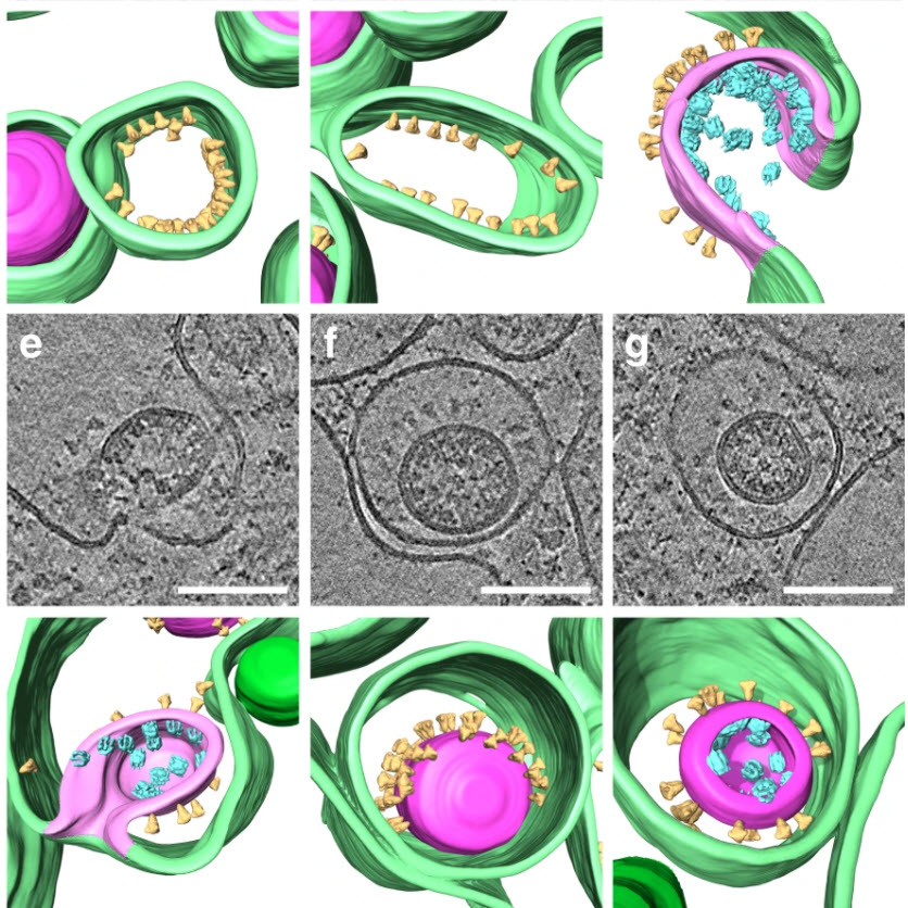
SARS-CoV-2 structure and replication characterized by in situ cryo-electron tomography
Severe acute respiratory syndrome coronavirus 2 (SARS-CoV-2), the causative agent of the COVID19 pandemic, is a highly pathogenic β-coronavirus. As other coronaviruses, SARS-CoV-2 is enveloped, replicates in the cytoplasm and assembles at intracellular membranes. Here, we structurally characterize the viral replication compartment and report critical insights into the budding mechanism of the virus, and the structure of extracellular virions close to their native state by in situ cryo-electr... Read more
Steffen Klein, Mirko Cortese, Sophie L. Winter, Moritz Wachsmuth-Melm, Christopher J. Neufeldt, Berati Cerikan, Megan L. Stanifer, Steeve Boulant, Ralf Bartenschlager, Petr Chlanda
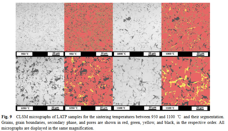
Lithium aluminum titanium phosphate (LATP) is one of the materials under consideration as an electrolyte in future all-solid-state lithium-ion batteries. In ceramic processing, the presence of secondary phases and porosity play an important role. In a presence of more than one secondary phase and pores, image analysis must tackle the difficulties about distinguishing between these microstructural features. In this study, we study the phase evolution of LATP ceramics sintered at temperatures b... Read more
Deniz Cihan GUNDUZ, Roland SCHIERHOLZ, Shicheng YUa, Hermann TEMPEL, Hans KUNGL, Rüdiger-A. EICHEL
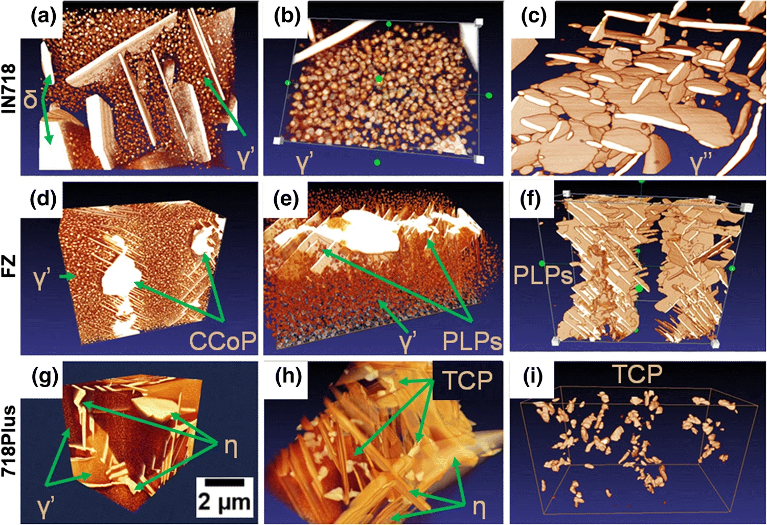
Inconel 718 (IN718) is the most popular precipitation-strengthened nickel-based superalloy introduced by the Huntington Alloys Division of INCO in 1959 (Ref Read more
Oskar Dziuba, Grzegorz Cempura, Agnieszka Wusatowska-Sarnek & Adam Kruk